Rib Cage Anatomy Male / Rib Cage Photos Media Storehouse - The thoracic cage is made up of bones and cartilage along, it consists of the 12 pairs of ribs with their costal cartilages and the.
Rib Cage Anatomy Male / Rib Cage Photos Media Storehouse - The thoracic cage is made up of bones and cartilage along, it consists of the 12 pairs of ribs with their costal cartilages and the.. At the chest, many rib bones connect to the sternum via costal cartilage,. The costal cartilages of the second through tenth ribs connect to the body of the sternum to form the bulk of the rib cage. It is made up of 12 pairs of ribs. The first rib is the widest, shortest and has the sharpest curve of all the ribs. Anatomy and physiology of human ribs.
Lesson by stan prokopenko in anatomy of the human body. They are strong enough to support the skeleton and protect the vital organs in the. The rib cage can be thought of as a handle on a bucket. The first rib is the widest, shortest and has the sharpest curve of all the ribs. Just like in the manubrium, slight concave indentations in the lateral sides of the body of the sternum provide stronger attachment points for the costal cartilages to prevent rib separation.
Untitled Human Figure Drawing Drawings Male Figure Drawing from i.pinimg.com Rib cage anatomy the rib cage, shaped in a mild cone shape and more flexible than most bone sets, is made up of varying elements such as the thoracic vertebra, 12 equally paired ribs, costal cartilage, and held together anteriorly by the sternum. Lesson by stan prokopenko in anatomy of the human body. Diagram of human body, liver rib cage, rib cage diagram labeled, rib cage diagram numbered, rib cage diaphragm, rib cage heart, rib cage organs anatomy, rib cage pain, stomach, diagram of human body, liver rib cage, rib cage diagram labeled, rib cage diagram numbered, rib cage diaphragm, rib cage. They are strong enough to support the skeleton and protect the vital organs in the. The volume of the rib cage is about 10% smaller in females than in males having the same height although the reason for this is presently unclear. Browse 6,946 human rib cage stock photos and images available, or search for human heart or skeleton to find more great stock photos and pictures. The upper edge is round and the lower sharp. Lateral view of a pair of ribs articulating with the thoracic vertebrae.
The spleen is used to filter red blood cells and hangs in the upper part of the abdomen.
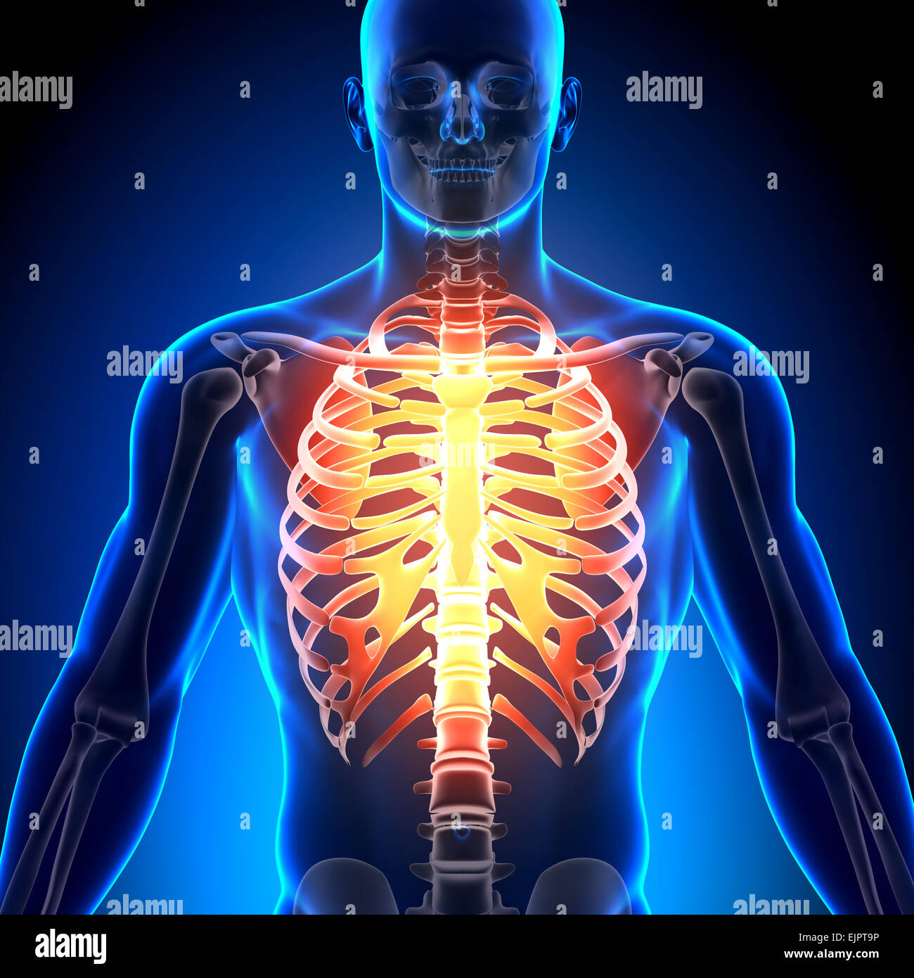
The superior surface is unique in that it is marked by two grooves that allow. It is made up of 12 pairs of ribs. A rib has a flat body, as you can see from the picture of the anatomy of the human rib cage. Just like in the manubrium, slight concave indentations in the lateral sides of the body of the sternum provide stronger attachment points for the costal cartilages to prevent rib separation. The bones of the rib cage are the sternum, the 12 thoracic vertebrae and the 12 pairs of ribs. The front edge ends with an ellipsoidal shape on which. This furrow isn't present in the 11th and 12th ribs. Although that is one key function, the ribcage does so much more. The rib cage can be thought of as a handle on a bucket. Lesson by stan prokopenko in anatomy of the human body. Doctors from medicinenet say that the middle and upper part of your spine contains 12 vertebrae that are attached to your rib cage. There are twelve (12) pairs of ribs and all articulate posteriorly with the thoracic vertebrae. The ribs are a set of twelve paired bones which form the protective 'cage' of the thorax.
The rib cage, or thoracic basket, consists of the 12 thoracic (chest) vertebrae, the 24 ribs, and the breastbone, or sternum. Normal anterior view of the rib cage and. The ribs are a set of twelve paired bones which form the protective 'cage' of the thorax. The cartilage that forms at the end of each rib (costal cartilage) attaches either. They articulate with the vertebral column posteriorly, and terminate anteriorly as cartilage (known as costal cartilage).
Print Of Medical Illustration Of Male Chest With Arteries Veins Heart And Rib Cage In 2021 Medical Illustration Male Chest Human Bones from i.pinimg.com Rib cage anatomy the rib cage, shaped in a mild cone shape and more flexible than most bone sets, is made up of varying elements such as the thoracic vertebra, 12 equally paired ribs, costal cartilage, and held together anteriorly by the sternum. Browse 6,946 human rib cage stock photos and images available, or search for human heart or skeleton to find more great stock photos and pictures. In 23 m … sexual dimorphism of human ribs The cartilage that forms at the end of each rib (costal cartilage) attaches either. There are twelve (12) pairs of ribs and all articulate posteriorly with the thoracic vertebrae. The spleen is used to filter red blood cells and hangs in the upper part of the abdomen. The volume of the rib cage is about 10% smaller in females than in males having the same height although the reason for this is presently unclear. The top edge of the manubrium has a depression called the suprasternal or jugular notch.
The sternum is a flat bone that is made up of three parts, the (1) manubrium, (2) body, and the (3) xiphoid process.
As in the typical ribs, the tubercle has a facet for articulation with the transverse process of vertebrae. The ribs are attached to the breastbone, which is the. The thoracic back describes the area of your back from just below the base of your skull to about 5 inches below the lower part of your shoulder blades. Abdominal muscle diagram 12 photos of the abdominal muscle diagram abdominal muscle anatomy bodybuilding, abdominal muscle diagram female, abdominal muscle groups diagram, human abdominal muscle diagram, lower abdominal muscle diagram, human muscles, abdominal muscle anatomy bodybuilding, abdominal muscle diagram female. Both men and women have 12 pairs of ribs.1 these ribs extend from the vertebrae to form the wall of the thoracic cavity (where the lungs and heart reside). Diagram of human body, liver rib cage, rib cage diagram labeled, rib cage diagram numbered, rib cage diaphragm, rib cage heart, rib cage organs anatomy, rib cage pain, stomach, diagram of human body, liver rib cage, rib cage diagram labeled, rib cage diagram numbered, rib cage diaphragm, rib cage. The sternum is a flat bone that is made up of three parts, the (1) manubrium, (2) body, and the (3) xiphoid process. A rib has a flat body, as you can see from the picture of the anatomy of the human rib cage. The primary causes of pain under the left rib cage. The head only articulates with the body of the t1 vertebra and therefore only one articulatory surface is present. Just like in the manubrium, slight concave indentations in the lateral sides of the body of the sternum provide stronger attachment points for the costal cartilages to prevent rib separation. It is made up of 12 pairs of ribs. There are twelve (12) pairs of ribs and all articulate posteriorly with the thoracic vertebrae.
The upper edge is round and the lower sharp. The exception to this anatomy rule are people born with specific genetic anomalies. Anatomy and physiology of human ribs. The superior surface is unique in that it is marked by two grooves that allow. 16 photos of the rib cage diagram with organs.
Male Rib Cage Anatomy Bones Stock Photo Alamy from c8.alamy.com This furrow isn't present in the 11th and 12th ribs. The upper edge is round and the lower sharp. The cartilage that forms at the end of each rib (costal cartilage) attaches either. The costal cartilages of the second through tenth ribs connect to the body of the sternum to form the bulk of the rib cage. The ribs are attached to the breastbone, which is the. Although that is one key function, the ribcage does so much more. As part of the bony thorax, the ribs protect the internal thoracic organs. In this video we discuss the structure of the rib cage or thoracic cage.
The primary causes of pain under the left rib cage.
Related posts of muscle anatomy rib cage abdominal muscle diagram. As part of the bony thorax, the ribs protect the internal thoracic organs. An enlarged or ruptured spleen can cause sudden or chronic pain under the left rib cage that ends up migrating towards the back and/or shoulders. A rib has a flat body, as you can see from the picture of the anatomy of the human rib cage. Although that is one key function, the ribcage does so much more. Each pair is numbered based on their attachment to the sternum, a bony process at the front of the rib cage which serves as an anchor point. The thoracic back describes the area of your back from just below the base of your skull to about 5 inches below the lower part of your shoulder blades. Lesson by stan prokopenko in anatomy of the human body. The bones of the rib cage are the sternum, the 12 thoracic vertebrae and the 12 pairs of ribs. As in the typical ribs, the tubercle has a facet for articulation with the transverse process of vertebrae. The top edge of the manubrium has a depression called the suprasternal or jugular notch. The ribs are a set of twelve paired bones which form the protective 'cage' of the thorax. The first seven pairs of ribs are called true ribs and connect to the sternum.


Posting Komentar
0 Komentar